
Live-Cell Imaging and Analysis System: Incucyte® SX1
Our most economical choice for live-cell imaging and analysis. Designed with the small lab in mind, the Incucyte® SX1 Live-Cell Analysis System allows you to see what your cells are doing and when, all from the stable environment of a tissue culture incubator. Leverage up to two total fluorescence channels simultaneously for long-term time-lapse experiments.
Enjoy walk-away convenience as images are automatically acquired and analyzed in a variety of formats. Insight, accuracy and reliability is available at every step in the process.
Key Features:
- Robust application suite – More than 25 validated applications for 2D and 3D cell cultures
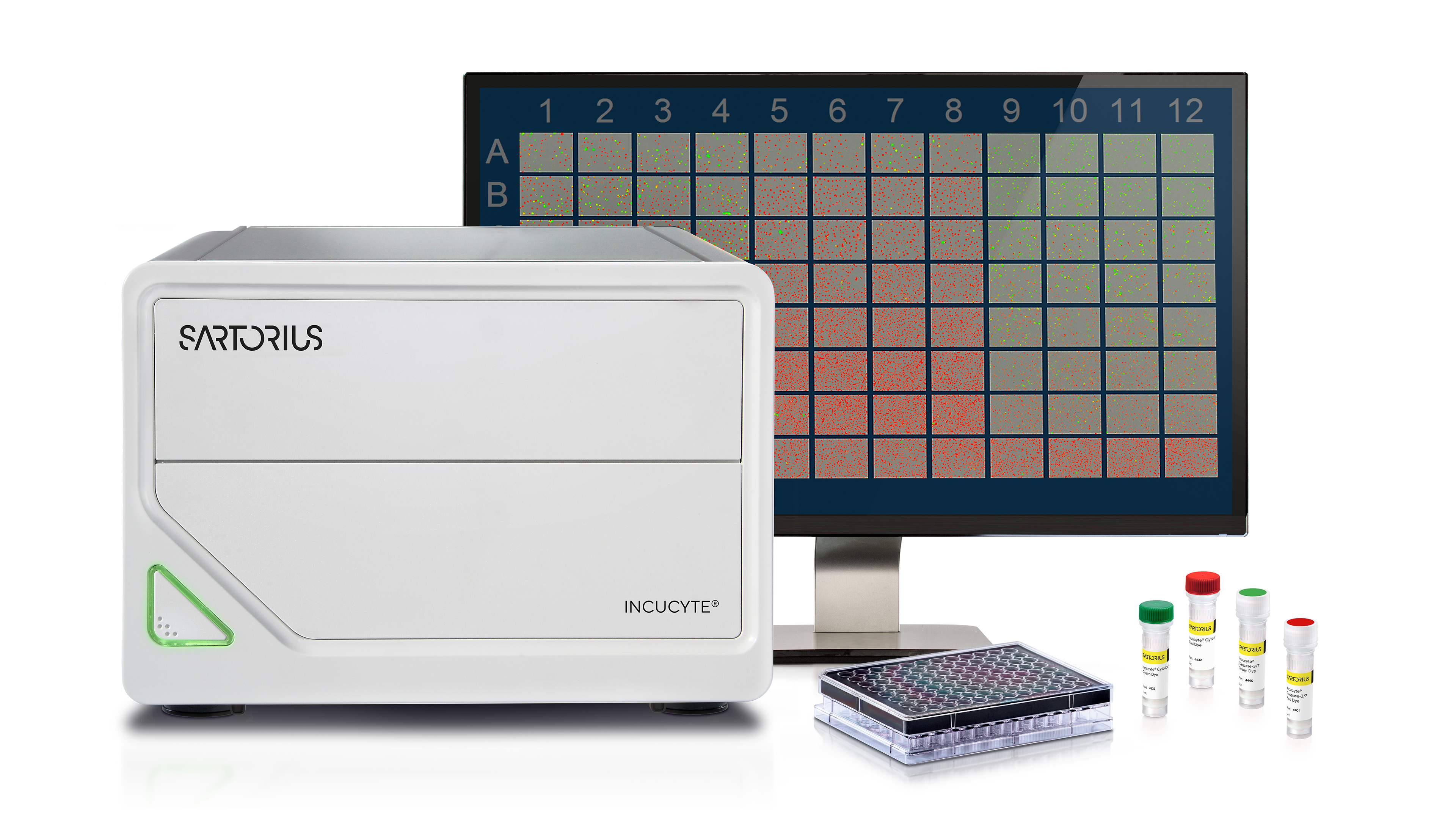
- Two-color Green/Red fluorescence plus HD Phase imaging
- 10X objective single position use – 4X or 20X objectives available separately
- Interchangeable tray to support over 700 vessels, up to two microplates in parallel
The Incucyte® SX1 Live-Cell Analysis System provides remote network capability and unlimited free user licenses to allow access to your cells 24 hours a day. Keep an eye on your budget and fast track your research with automation at every step!
Explore the Incucyte® SX1


See what the SX1 has to offer in Live-Cell Imaging & Analysis
Feel Empowered with Incucyte® SX1
Key Features

Simple, Flexible Sample Prep
- Compatible with a wide range of applications and culture vessels
- Maximize efficiency while reducing artifacts with Incucyte® Reagents for live-cell assays

Set Up & Walk Away
- Streamlined experimental set up
- Remote, networked access

Acquire and View Images Over Time
- Fluorescence and HD phase imaging modes
- Minimize cell disturbance with a mobile optical train

Analyze in Real-Time
- Efficient and reproducible image analysis
- Powerful visualization of images and kinetic measurements
Case Studies & Testimonials
KOL Interview: Dr. Clint Gray & Raka Mitra Pioneering a 3D Model for Infant Vascular Tumors
Infant vascular tumour research has been a main focus for 2D research... until now. Find out more on Raka Mitra, the individual who built the first 3D model for infant vascular tumour analysis.
In this exclusive interview with both Dr. Clint Gray, Director of GMRI and Raka Mitra, dive into the depths of their transformative research supported by the Incucyte® SX1.
Incucyte® SX1 Applications
Incucyte®'s live-cell applications provide real-time insight into morphological and phenotypic changes for pathway and mechanistic studies by observing time and cell dependent treatment effects. Capable of working with rare or expensive cell types, you can validate cell morphology and phenotype at all stages of cell maintenance and modification, and analyze a wide range of phenotypic cell-based assays. With Incucyte®, you can make informed decisions, optimize workflows, and accelerate your next discovery.
Accelerate your next discovery with Incucyte’s suite of live-cell applications.


Cell Monitoring & Workflows
Imaging Vessels
The Incucyte® Live-Cell Analysis Systems are compatible with a wide range of culture vessels – currently over 700 and growing! From 6-well to 384-well microtiter plates to standard tissue culture flasks, the Incucyte® can support your live-cell imaging and analysis needs.
SX1 Specifications
Category | Attributes | Description |
|---|---|---|
Optical System | Fluorescence Channels | 2 |
Optical Module | Green/Red | |
Label-Free HD Phase Imaging | ||
Objectives | 10X (included) 4X (available for purchase) 20X (available for purchase | |
Objective Automation | Manual swap | |
Multi-User Support | Remote Network Capability Unlimited, Free user licenses | |
Incubation | Incubator Size | >200 L required |
Incubation to 42°C | ||
Software Module Compatibility | Incucyte® Cell-by-Cell Incucyte® Spheroid Incucyte® Cell Migration Incucyte® Chemotaxis Incucyte® Neurotrack Incucyte® Angiogenesis Incucyte® AI Cell Health | |
Capacity and Vesselware Compatibility | Interchangeable Trays | 1 |
Microplate Capacity | 2 | |
Microplate Compatibility | 6, 12, 24, 48, 96, 384-well | |
Flask Compatibility | T-25, T-50, T-75, T-100, T-175 | |
Other Labware Compatibility | 35mm, 60mm, 100mm, 150mm dishes and chamber slides | |
Computing and Storage | Operating System | 64-bit Windows 10 |
Controller Data Storage | 27.3 TB | |
Expandable RAID Storage | Add an additional 32.7 TB of storage with Incustore | |
Controller Memory | 64 GB |
Related Products


SX1 Resources
Featured Resources


FAQ for Live-Cell Imaging and Analysis Instrument: Incucyte® SX1
Fluorescent channels vary depending on the instrument. In the case of the Incucyte® SX1 is equipped with two-color Green/Red fluorescence plus HD Phase imaging.
The Incucyte® itself is not an incubator but sits within a standard tissue culture incubator so cells are maintained in a physiologically relevant environment. For the Incucyte® SX1, an incubator larger than 200L is recommended.
The Incucyte® SX1 is a great budget-friendly option that can support the same applications as an Incucyte® S3, but at a lower throughput for smaller labs with less users. It is an ideal option for labs who are looking for a model that better suits their budget. In addition, the Incucyte® SX1 can be upgraded to an Incucyte® S3 if the additional features and an increased throughput is needed in the future.
What to Expect from our Virtual Demo
Because we want to provide the best and most relevant support to your research, Sartorius experts are available to demonstrate the features and benefits of our live-cell imaging and analysis systems in a personalized manner.
- Initial consultation with your local support team to understand your interests
- Virtual demo at no cost to you!
- Total demo time would be 2-4 hours, depending on number of applications
See how the Incucyte® can support your research needs, from setting up an assay to analyzing and exporting your data.