What is Antibody Internalization?
Antigen-mediated antibody internalization plays an important role in several antibody-based therapeutics. There is a continued growing body of interest in the application of antibodies as therapeutic agents for delivery of treatments to sites or cells of interest with promise of increased efficacy and reduced side effect potential. The significance and impact of therapeutic antibodies as effective treatments for disease span across numerous areas such as autoimmunity, cancer and inflammation, cardiovascular and infectious diseases, and many other disorders.
Current Research and Therapeutic Landscape
The evolving therapeutic impact of antibody internalization on the antibody discovery field and targeted-mediated internalization includes the delivery of highly toxic drugs to cancer cells via antibody drug conjugates (ADCs), removal or degradation of surface receptors from cancer cells (i.e. EGFR), and antibody immunotherapies used to identify tumor cells for immune cell killing (i.e. ADCC or ADCP). Additionally, measuring and optimizing functional responses to antibodies, such as antibody clearance is of importance. Pinocytosis, which is one of the main elimination routes of antibodies, requires antibody optimization for qualitative pharmacokinetic measurements during antibody development.
As each approach requires a series of antibody features, for example, to enable maintenance on the cell surface for identification of tumor cells, or for rapid internalization when delivering ADC’s, it is important to understand the uptake profile and clearance of antibody candidates for optimal antibody engineering and internalization characteristics.
Expanding and Improving Existing Methods
Current approaches are limited by requiring labor-intensive labeling of each antibody, washing steps, and/or single-time point analysis. This can lead to unnecessary and lengthy processes, loss of cells, or missed and incorrect measurements due to limitations of single time-point analysis. Therefore, throughout the entire antibody screening and functional characterization process, there is a strong need to have an efficient, affordable method to fully and rapidly quantify antibody internalization using real time live-cell analysis.
The Solution
Sartorius is proud to offer a new, novel solution that is compatible with human, mouse and rat test antibodies (utilizing Incucyte® Human, Mouse IgG1, or Rat Fabfluor-pH Red Antibody Labeling Reagent or Incucyte® Human Fabfluor-pH Orange Antibody Labeling Reagent) for rapid, kinetic, high-throughput analysis of antibody internalization.
Application
Introducing Incucyte® Antibody Internalization Assays
An integrated solution to automatically visualize and quantify antibody internalization, in real time and in 96-well formats— inside your tissue culture incubator.
(Video) Kinetic monitoring of antibody internalization with the Incucyte® Live-Cell Analysis System and Incucyte® Fabfluor-pH Antibody Labeling Reagent.
Herceptin shows a positive response (as measured by red fluorescence) over 12 hours in BT-474 cells. IgG isotype used as a negative control.
Download the Incucyte® Fabfluor-pH Antibody Labeling Reagents Product Guide
Key Advantages
Key Advantages of Incucyte® Antibody Internalization Assays
- Combine Sensitive, Kinetic Fluorescent Measurement - of internalization with images and movies for visual confirmations
- Rapid, Single-Step Labeling - paired with mix-and-read protocols for efficient testing of antibody panels
- High-Throughput, Reproducible - antibody screening data to provide crucial functional insight and unlock your Antibody Discovery potential
Did you know?
The all-new Incucyte integrated software offers unparalleled ease of use and the capability for live-cell imaging and analysis. Learn More
Easily visualize and measure antibody internalization over time using Incucyte® Fabfluor-pH Antibody Labeling Reagent and automated analysis tools.
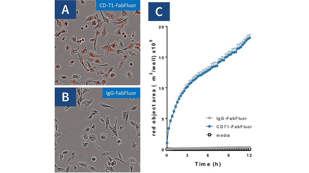
HT1080 cells were treated with either Incucyte® Fabfluor-pH labeled α-CD71 or IgG1 isotype control (4 µg/mL), HD phase and red fluorescence images were captured every 30 minutes over 12 hours using a 10x magnification. Images of cells treated with Fabfluor-α-CD71 display red fluorescence in the cytoplasm (A). Cells treated with labelled isotype control display no cellular fluorescence (B). Red object area over time is generated automatically, showing rapid increase in cells treated with labeled α-CD71 and no signal with isotype control (C). Images shown taken at 6 hours post treatment, data shown as mean of 3 wells ± SEM.

Distinguish differences in internalization responses across multiple cell types driven by cell surface antigen expression.
Jurkat (T cell like) and Raji (B cell like) cells (30K/well) were treated with various Incucyte® Fabfluor-pH labeled antibodies (4 µg/mL) to show the specific nature. HD phase and red fluorescence images were captured every 30 minutes using a 20x objective over 12 hours. Time course data (A & B) demonstrates a response profile in both cell lines that matches the expected expression profile of the tested markers, and confirms that an internalization response is only present when the specific antigen is expressed. Note that the assay can distinguish differences in response of Raji cells (steeper response curve for α-CD71 and therefore faster rate of internalization) compared to Jurkat cells. The area under the curve (AUC) analysis demonstrates the overall profile in both cell lines (C). All data shown as a mean of at least 4 wells ± SEM, time course data shown as normalized red area.

Reveal concentration-dependent responses and conduct pharmacological analyses.
BT-474 Her2 positive cells were treated with increasing concentrations of Fabfluor-pH labeled Herceptin using a simple mix-and-read protocol. The time course graph displays an increase normalized red area over time with increasing Herceptin concentrations (A); Area under the curve analysis of this response displays a clear concentration dependent response with an EC50 of 323 ng/mL (B). All data shown as a mean of 3 wells SEM, time course data shown as normalized red area.
Rapid, Single Step Labeling Paired With Mix-and-Read Protocols for Efficient Testing of Antibody Panels
Rapidly label multiple test antibodies and then simply mix and read to measure antibody internalization. No labeling optimization or wash step required. Labeling of a panel of antibodies can be achieved in under 15 minutes.

High-Throughput, Reproducible Antibody Screening Data to Provide Crucial Functional Insights and Unlock Your Antibody Discovery Potential
Screen large numbers of antibodies in 96- or 384-well format to generate consistent, cell-specific internalization response data to fully characterize test antibodies.
Achieve robust functional antibody screening with Incucyte® Fabfluor-pH labeled antibodies.
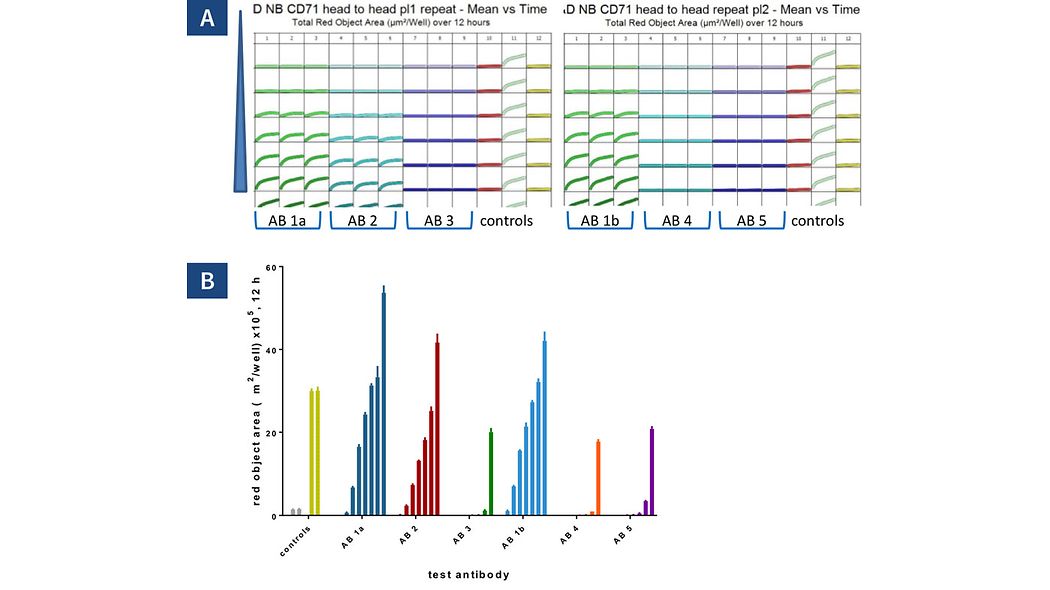
Six different CD71 antibodies including one clone from 2 different suppliers (clone 1a & 1b) were tested head to head in HT1080 cells. The antibodies were labeled in a single step with Incucyte® Fabfluor-pH Antibody Labeling Reagent prior to addition to cells and the internalization signal captured every 30 minutes over 12 hours using a 10x magnification. Plate views taken from Incucyte show clear positive and negative control responses in column 11 & 12 with concentration dependent responses for each antibody across two plates (A). Control responses at 12 hours display a clear positive response generating Z’ values of 0.86 and 0.77 (B). Head to head analysis of antibody data shows a range of responses across these clones (B). All data shown as mean of 3 wells +SEM, controls shown as mean of 8 wells.
Commonly Used Assays
| Incucyte® | Flow Cytometry | Fluorescence Microscopy | |
|---|---|---|---|
| Biological Insight | |||
| Real-time cell visualization |
| ||
| High sensitivity |
| ||
| Rate measurements | |||
| Productivity | |||
Integrated analysis |
| ||
Mix & Read Protocol |
| ||
96-well throughput |
|
| Incucyte® Fabfluor-pH | Top Competitor | Direct Labeling | |
|---|---|---|---|
Real-time cell visualization |
| ||
High sensitivity | |||
Rate measurements |
Table 1.
Comparison between Incucyte® Live-Cell Analysis System versus Traditional methods and Incucyte® Fabfluor-pH versus other reagents and Direct Label Methods; highlighting Incucyte® advantages.
Ordering Information
Ordering Information
Product | Qty. | Cat. No. |
|---|---|---|
| Incucyte® Human Fabfluor-pH Red Antibody Labeling Reagent | 1 vial (50 µg) | |
| Incucyte® Mouse IgG1 Fabfluor-pH Red Antibody Labeling Reagent | 1 vial (50 µg) | |
| Incucyte® Rat Fabfluor-pH Red Antibody Labeling Reagent | 1 vial (50 µg) | |
| Incucyte® Mouse IgG2a Fabfluor-pH Red Antibody Labeling Reagent | 1 vial (50 µg) | |
| Incucyte® Mouse IgG2b Fabfluor-pH Red Antibody Labeling Reagent | 1 vial (50 µg) | |
Incucyte® Human Fabfluor-pH Orange Antibody Labeling Reagent | 1 vial (50 µg) |